
Elbow Dysplasia
This page is for educational purposes only and is not a substitute for veterinary diagnosis or emergency care. Always consult your primary veterinarian or a rehabilitation veterinarian before starting treatment. If your pet cannot walk, has sudden paralysis, severe pain, or breathing difficulty, seek urgent veterinary attention.
What is Elbow Dysplasia?
Also known as: developmental elbow disease; canine elbow dysplasia (CED); medial compartment disease.
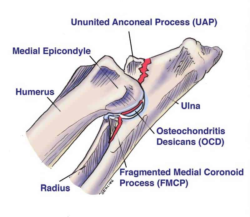
“Elbow dysplasia” is an umbrella term for developmental abnormalities of the elbow: fragmented medial coronoid process, ununited anconeal process, osteochondritis dissecans of the medial humeral condyle, and joint incongruity. These lesions produce inflammation, cartilage injury, and early osteoarthritis. Dogs — especially medium, large, and giant breeds — are primarily affected; cats more often have elbow OA from other causes, though developmental issues can occur.
Clinical signs often begin in adolescence: forelimb lameness, elbow thickening, and exercise intolerance. Advanced imaging is frequently required for complete lesion mapping.
Treatment may include arthroscopy, osteotomy strategies, medical OA management, and weight control. Rehabilitation is a long-term partner for comfort, muscle, and activity pacing whether or not surgery is performed.
Common signs to watch for
Signs vary by severity and by whether your pet is a dog or cat. Owners of dogs and cats often notice:
- Forelimb lameness (uni- or bilateral), sometimes subtle or shifting
- Elbow swelling; reluctance to flex fully
- Outward turning of the foot or elbow abduction at stance
- Stiffness after rest; worse after heavy play
- Reduced willingness to jump down from heights
Causes & contributing factors
- Genetic developmental disorders of elbow formation and congruence
- Osteochondrosis-spectrum disease within the elbow
- Growth-related incongruity between radius and ulna
- Excess body weight amplifying joint load in a vulnerable elbow
How veterinary rehabilitation helps
Rehab programmes target pain relief, maintain motion, and strengthen the kinetic chain from paw to scapula and core so the dysplastic elbow is better supported.
Post-operative and conservative pathways both use graded therapeutic exercise; hydrotherapy may reduce impact while preserving fitness when cleared.
Owner education on lifelong OA management — surfaces, weight, and flare plans — is central.
Rehabilitation plans at RehabVet are individualised after a veterinary assessment. We coordinate with your primary vet when imaging, medication, or surgery is part of the overall plan.
Modalities & services commonly used at RehabVet
Depending on your pet’s examination findings, comfort, and goals, a plan may include one or more of the following:
Expected rehabilitation goals
Goals are set for the individual patient. Typical aims may include (not guarantees — outcomes vary):
- Improve forelimb comfort and use
- Preserve functional elbow range
- Build supporting musculature and gait quality
- Support healthy body weight
- Provide a sustainable home and clinic exercise plan
We do not publish invented success percentages. Progress is tracked clinically (gait, strength, range of motion, pain behaviours, and home function) and plans are adjusted over time.
When to seek veterinary care
- Persistent forelimb limp in a growing dog
- Elbow swelling or reduced motion
- Bilateral forelimb lameness or declining play
- Known breed risk with early mobility changes — discuss screening with your vet
Medial coronoid disease / FMCP is among the most frequently diagnosed components, but many elbows have combined pathology. Imaging clarifies the individual case.
Responsible breeding and screening reduce risk at a population level. For an individual puppy, appropriate nutrition, controlled growth, and avoiding excessive impact help but cannot erase genetic risk.
Yes. Many dysplastic elbows already show OA at diagnosis. Rehab focuses on comfort, strength, and function within that reality — coordinated with your vet’s medical plan.
Related reading & patient stories
Book a rehabilitation assessment
If your pet has been diagnosed with Elbow dysplasia, or you are noticing mobility changes, our team can assess and design a multimodal rehab plan.
Educational content only — not a diagnosis. For emergencies, contact your nearest veterinary hospital.
Related Conditions
- ConditionAngular Limb DeformityAngular limb deformity describes abnormal angulation or rotation of a limb from asymmetric growth-plate function or trauma, altering joint alignment and gait in dogs.Learn more
- ConditionHip DysplasiaCanine hip dysplasia is a developmental malformation of the hip joint that leads to laxity, pain, and secondary osteoarthritis — managed with multimodal care including physiotherapy and hydrotherapy.Learn more
- ConditionLegg-Calvé-Perthes DiseaseLegg-Calvé-Perthes disease is avascular necrosis of the femoral head in young small-breed dogs, causing hip pain, lameness, and collapse of the femoral head.Learn more