
Intervertebral Disc Disease (IVDD)
This page is for educational purposes only and is not a substitute for veterinary diagnosis or emergency care. Always consult your primary veterinarian or a rehabilitation veterinarian before starting treatment. If your pet cannot walk, has sudden paralysis, severe pain, or breathing difficulty, seek urgent veterinary attention.
What is Intervertebral Disc Disease (IVDD)?
Also known as: slipped disc; herniated disc; disc disease; Hansen Type I / Type II disc disease.
Intervertebral discs sit between most vertebrae and act as shock absorbers. Each disc has a tough outer annulus and a softer inner nucleus. In IVDD, disc material degenerates and may protrude or extrude into the spinal canal, compressing the spinal cord (myelopathy) and/or nerve roots (radiculopathy). Dogs are far more commonly affected than cats.
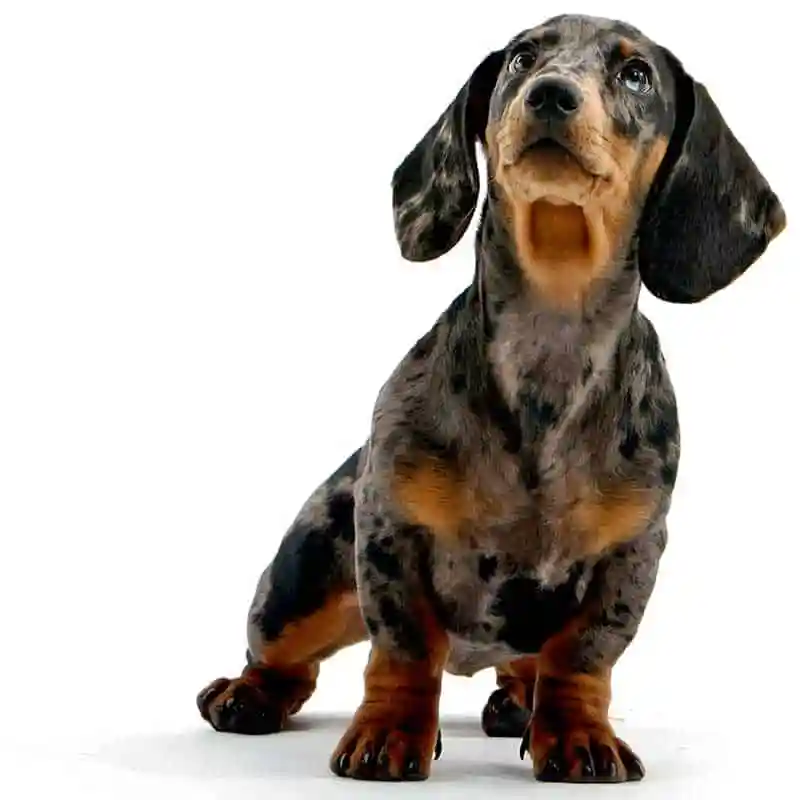
Two classic patterns are described. Hansen Type I tends to present more acutely in chondrodystrophic breeds (for example Dachshunds, French Bulldogs, Beagles, Shih Tzus) when the nucleus hardens and can extrude suddenly. Hansen Type II tends to involve gradual annular bulging in larger, non-chondrodystrophic breeds. A third pattern — acute non-compressive or “high-velocity, low-volume” disc extrusion — can follow strenuous activity and may not leave ongoing compression.
Severity ranges from pain and reluctance to jump, through knuckling and a wobbly gait, to inability to walk or loss of deep pain sensation. Diagnosis and grading belong with your veterinarian (often including advanced imaging). Rehabilitation does not replace emergency assessment, surgery when indicated, or medical management — it supports comfort, neuro-recovery, and safe return to function alongside that care.
Common signs to watch for
Signs vary by severity and by whether your pet is a dog or cat. Owners of dogs often notice:
- Reluctance to jump, climb stairs, or play as usual
- Crying out, hunched posture, tense back or neck muscles
- Hind-limb weakness, knuckling, crossing, or a drunken (ataxic) gait
- Dragging paws, difficulty rising, or inability to walk
- Reduced appetite or activity; holding the head low with neck pain
- In severe cases: paralysis and changes in bladder or bowel control
Causes & contributing factors
- Age-related and breed-related disc degeneration (chondrodystrophic conformation)
- Acute extrusion during activity in a disc already weakened by degeneration
- Gradual disc protrusion (Type II) in larger breeds
- Trauma can precipitate rupture of a compromised disc but is not the same as degenerative IVDD alone
- Obesity and high-impact repetitive loading may increase mechanical stress on the spine
How veterinary rehabilitation helps
After veterinary diagnosis and clearance, rehab aims to reduce pain behaviours, protect the spine during healing, and rebuild controlled movement. Early phases often emphasise careful handling, appropriate rest or controlled activity, and modalities that support comfort without forcing neurological recovery.
As pain settles and neurological status allows, plans typically progress to assisted standing, proprioceptive work, therapeutic exercise, and — when appropriate — hydrotherapy or underwater treadmill training to rebuild strength with reduced load. Home programmes help owners manage toileting, sling support, and safe surfaces.
For surgical and non-surgical pathways alike, rehab is individualised. Progress is tracked by gait quality, strength, pain signs, and daily function — not by promised percentages.
Rehabilitation plans at RehabVet are individualised after a veterinary assessment. We coordinate with your primary vet when imaging, medication, or surgery is part of the overall plan.
Modalities & services commonly used at RehabVet
Depending on your pet’s examination findings, comfort, and goals, a plan may include one or more of the following:
Expected rehabilitation goals
Goals are set for the individual patient. Typical aims may include (not guarantees — outcomes vary):
- Improve comfort and reduce protective muscle guarding around the spine
- Support return of voluntary motor control and coordinated gait when neurologically possible
- Rebuild core and limb strength with spine-safe loading
- Teach owners safe handling, restriction, and toileting support
- Reduce compensatory strain in shoulders, elbows, and hips from altered gait
We do not publish invented success percentages. Progress is tracked clinically (gait, strength, range of motion, pain behaviours, and home function) and plans are adjusted over time.
When to seek veterinary care
- Sudden inability to walk, knuckling, or collapse — seek urgent veterinary care
- Severe pain, crying when touched, or refusal to move the neck or back
- Loss of bladder or bowel control, or a bloated/uncomfortable abdomen
- Rapidly worsening weakness over hours to days
- Any suspicion of spinal disease before starting exercise, hydrotherapy, or massage at home
No. Treatment depends on neurological grade, pain level, imaging findings, and how quickly signs progress. Some dogs are managed medically with strict rest and pain control; others need surgical decompression. Your primary vet or neurologist decides the pathway. Rehabilitation can support both surgical and non-surgical recovery once cleared.
Timing depends on whether your dog is awaiting imaging, resting medically, or recovering from surgery. Gentle, supervised rehab may begin early for pain and positioning, while loading and hydrotherapy wait until your vet confirms it is safe. Never start intensive exercise during acute spinal compression without veterinary guidance.
IVDD is much less common in cats than dogs, but disc disease and spinal pain can occur. Cats more often show reduced jumping, hiding, or irritability rather than dramatic hind-limb paralysis. See our guide to neurological conditions in cats for the feline angle, or book a cat-focused assessment.
Recovery potential varies with severity, time to treatment, deep pain status, and individual factors. Many dogs improve meaningfully with appropriate medical or surgical care plus rehab; others need long-term mobility aids. We avoid promising outcomes — we measure progress clinically and adjust the plan with your vet.
Related reading & patient stories
Book a rehabilitation assessment
If your pet has been diagnosed with IVDD, or you are noticing mobility changes, our team can assess and design a multimodal rehab plan.
Educational content only — not a diagnosis. For emergencies, contact your nearest veterinary hospital.
Related Conditions
- ConditionWobbler SyndromeWobbler syndrome (cervical spondylomyelopathy) refers to cervical spinal cord compression in dogs, producing a characteristic wobbly gait, neck pain, and progressive neurological deficits.Learn more
- ConditionDiscospondylitisDiscospondylitis is infection of an intervertebral disc and neighbouring vertebral endplates, causing spinal pain, stiffness, fever, and sometimes neurological deficits in dogs (rarely cats).Learn more
- ConditionFibrocartilaginous Embolism (FCE)Fibrocartilaginous embolism (FCE) is an acute spinal cord infarction caused by disc material blocking a spinal blood vessel — typically sudden, often asymmetric, and usually non-progressive after the first hours.Learn more