
Luxating Patella
This page is for educational purposes only and is not a substitute for veterinary diagnosis or emergency care. Always consult your primary veterinarian or a rehabilitation veterinarian before starting treatment. If your pet cannot walk, has sudden paralysis, severe pain, or breathing difficulty, seek urgent veterinary attention.
What is Luxating Patella?
Also known as: patellar luxation; slipped kneecap; MPL (medial patellar luxation); kneecap dislocation.
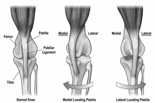
The patella normally tracks in the trochlear groove of the femur. In luxating patella, conformational factors — shallow groove, malalignment of the quadriceps mechanism, tibial tuberosity position, and limb deformity — allow the patella to displace, most often medially in small breeds and sometimes laterally in larger dogs.
Owners often see intermittent “skipping” or holding up a hind leg for a few steps until the patella reduces. Grades I–IV describe severity from intermittent manual luxation to permanent luxation with limb deformity. Concurrent cranial cruciate disease is not rare in affected stifles.
Mild grades may be managed with weight control, physiotherapy, and activity modification; higher grades or painful dysfunction often need surgical realignment. Rehabilitation supports muscle balance, gait, and post-operative recovery.
Common signs to watch for
Signs vary by severity and by whether your pet is a dog or cat. Owners of dogs often notice:
- Intermittent skipping or hopping on a hind limb
- Occasional cries when the kneecap luxates or reduces
- Bow-legged (genu varum) appearance in chronic medial luxation
- Reluctance to jump; stiffness after rest
- Progressive lameness if osteoarthritis or cruciate disease develops
Causes & contributing factors
- Congenital/developmental malalignment of the extensor mechanism
- Shallow femoral trochlear groove
- Breed predisposition (many toy and small breeds; some large breeds)
- Trauma (less common primary cause than developmental luxation)
- Secondary changes from limb deformity or muscle imbalance
How veterinary rehabilitation helps
For lower-grade luxation, rehab emphasises quadriceps and hip abductor/adductor balance, core stability, proprioception, and controlled strengthening to improve tracking and confidence.
After surgical correction, programmes follow surgeon protocols for swelling, range, weight-bearing, and progressive exercise. Hydrotherapy may assist gait retraining when cleared.
Owners learn which games to avoid (high-torque twisting), how to monitor skipping frequency, and when to seek recheck.
Rehabilitation plans at RehabVet are individualised after a veterinary assessment. We coordinate with your primary vet when imaging, medication, or surgery is part of the overall plan.
Modalities & services commonly used at RehabVet
Depending on your pet’s examination findings, comfort, and goals, a plan may include one or more of the following:
Expected rehabilitation goals
Goals are set for the individual patient. Typical aims may include (not guarantees — outcomes vary):
- Reduce pain and frequency of clinically problematic luxation episodes
- Improve hind-limb strength and neuromuscular control
- Support post-operative healing and gait normalisation when surgery is performed
- Maintain healthy body weight to unload the stifle
- Limit secondary osteoarthritis progression through smart activity
We do not publish invented success percentages. Progress is tracked clinically (gait, strength, range of motion, pain behaviours, and home function) and plans are adjusted over time.
When to seek veterinary care
- Frequent skipping, persistent limp, or pain associated with the kneecap
- Sudden worsening that could indicate cruciate ligament injury
- Young dogs with bilateral bowing and mobility limits
- Before breeding discussions in dogs with known luxation — veterinary advice
No. Grade, pain, function, and lifestyle guide decisions. Many grade I–II dogs do well with conservative care and rehab; higher grades or functional impairment more often need surgery.
Rehab cannot deepen a shallow groove or fully correct bony malalignment. It can improve muscle support and function, and it is essential after surgical realignment.
Patellar luxation occurs in cats but is less commonly discussed than in dogs. Any intermittent hind-limb skipping in a cat still warrants veterinary exam.
Yes — dogs with chronic patellar luxation can have altered stifle mechanics and concurrent cranial cruciate disease. Sudden new lameness needs prompt assessment.
Related reading & patient stories
Book a rehabilitation assessment
If your pet has been diagnosed with Luxating patella, or you are noticing mobility changes, our team can assess and design a multimodal rehab plan.
Educational content only — not a diagnosis. For emergencies, contact your nearest veterinary hospital.
Related Conditions
- ConditionAngular Limb DeformityAngular limb deformity describes abnormal angulation or rotation of a limb from asymmetric growth-plate function or trauma, altering joint alignment and gait in dogs.Learn more
- ConditionHip DysplasiaCanine hip dysplasia is a developmental malformation of the hip joint that leads to laxity, pain, and secondary osteoarthritis — managed with multimodal care including physiotherapy and hydrotherapy.Learn more
- ConditionElbow DysplasiaElbow dysplasia is a group of developmental elbow disorders — including FMCP, UAP, OCD, and incongruity — that cause forelimb pain and progressive osteoarthritis, mainly in dogs.Learn more