In memories of all immobile dogs that were not given a second chance
A fibrocartilaginous embolism, or FCE for short, is a blockage in a blood vessel in the spinal cord. It’s often referred to as a spinal cord stroke.
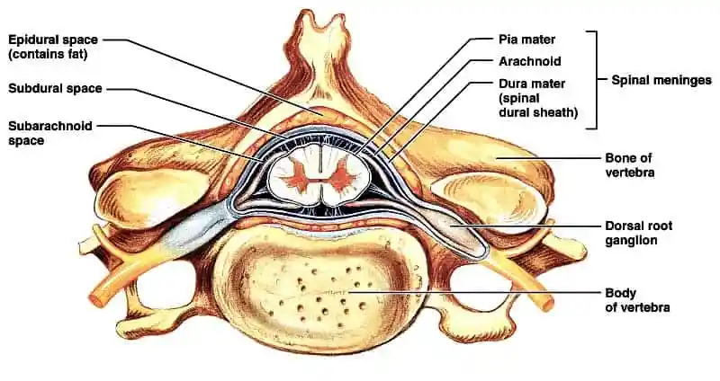
The vertebral column is made up of small bones called vertebrae that are joined together by intervertebral discs. The discs function as cushions between the vertebrae and allow the spine to flex. They are round in shape, fibrous on the outside, and contain a gel-like substance on the inside called the nucleus pulposus.
One of the jobs of the vertebral column is to protect the spinal cord inside it. The spinal cord is similar to a long cable of nerves that sends messages to and from the brain and regulates the body’s reflexes. The spinal cord is fed by a system of blood vessels.
DACVSMR, CCRP
Professor of Orthopedic Surgery & Director of Surgical Service
Robin Downing, DVM, MS, DAAPM, DACVSMR, CVPP, CCRP
Diplomate of the American Academy of Pain Management, is a a founder and past-president of the International Veterinary Academy of Pain Management.
Janet B. Van Dyke, DVM
Diplomate American College of Veterinary Sports Medicine and Rehabilitation, CCRT, CEO
Ludovica Dragone, DVM, CCRP
Vice President of VEPRA, Veterinary European of Physical Therapy and Rehabilitation Association.
Andrea L. Henderson, DVM, CCRT, CCRP
Resident, Canine Sports Medicine and Rehabilitation
MS, DVM, MBA, PhD
President Securos. Inc
Unfortunately, this is a question that we cannot answer. No dietary, environmental, or inherited factors, nor any underlying diseases have been identified that predispose to fibrocartilaginous embolism FCE in the majority of dogs. Miniature schnauzers with hyperlipidemia (an inherited tendency to develop high blood fat) and Shetland sheepdogs with hypothyroidism do seem to be predisposed to Fibrocartilagenous Embolism FCE; these dogs may have a slightly different condition from regular FCE.
A fibrocartilaginous embolism occurs when a fragment of the nucleus pulposus inside an intervertebral disc escapes into the blood vessel of the spinal cord and causes an obstruction. This affected area of the spinal cord then dies.
Unfortunately, neurologic loss that occurs within the first 24 hours is usually permanent. The good news is the condition isn’t progressive. Any pain usually resolves within 12 to 24 hours. And with immediate treatment, primarily involving very intensive physical therapy, most dogs experience significant recovery.
An FCE typically results from an injury to the spinal cord often caused when a dog jumps or lands awkwardly. Sometimes vigorous exercise can do it. Dog fights, really rough play, or any sort of accidental trauma can also lead to an FCE.
Fibrocartilaginous embolism are rarely seen in cats and occur most often in large and giant breed male dogs, and also miniature Schnauzers and Shelties between three and six years of age. It’s possible an underlying condition common in these breeds called hyperlipidemia, which is a high blood cholesterol level, could be a contributing factor in smaller dogs who acquire the condition.
Signs of a fibrocartilaginous embolism usually appear suddenly and follow a period of exercise or what otherwise seems like a mild injury or trauma.
Symptoms can include sudden, severe pain that makes the dog cry or yelp, followed by lessening pain after a few minutes or hours; signs of weakness; partial to full paralysis of a rear limb; a wobbly or uncoordinated gait; and lack of a pain response after initial signs of painfulness, yet the dog still can’t use his body normally.
Small dogs can:
You will need to provide a thorough history of your dog’s health leading up to the onset of symptoms, the type of activities your dog engages in, and any injuries that you suspect to have recently occurred. Your veterinarian will rule out other causes, such as spinal tumor, intervertebral disc disease, or fracture before settling on a diagnosis. The above mentioned conditioned are very painful, therefore, a lack of pain can be indicative of an embolism in the spinal cord. Keep in mind that though there may be a lack of pain, the condition can be progressive and may affect long-term damage to the spine and neurological system. Immediate and supportive care is essential.
Routine laboratory test results, such as urinalysis and complete blood counts, are usually unremarkable. A sample of cerebral spinal fluid (CSF) may be taken for analysis, and a sample of blood from the veins and arteries of the spinal cord may show microscopic fragments of fibrocartilage. Radiographic imaging studies may help in diagnosis. Apart from routine radiography, magnetic resonance imaging (MRI) remains the best diagnostic technique for viewing the spinal cord. In the later stage of fibrocartilaginous embolic myelopathy, swelling may be present at the site of the blockage.
As a non-surgical disease of the spinal cord, rehabilitation has a huge role to play in managing dogs diagnosed with FCE. Following the assessment of our animal rehabilitation practitioner, we will custom design a treatment plan unique for them. Maintenance of physical function and prevention of secondary compensatory changes is vital to your dog making good progress throughout the rehabilitation process. The level to which a patient is affected by the FCE will vary from dog to dog. Furthermore, the rate at which a patient progresses will dictate the necessary adjustments to the rehabilitation plan. Ongoing re evaluation is essential to allow an effective and appropriate treatment course.
In the initial stage following diagnosis, the aims are as follow:
Depending on your dogs progress, the aims might then progress to:
6 weeks on from diagnosis your chartered physiotherapist would aim to:
Correct positioning, massage and stretches can be implemented in the early days. sensory stimulation techniques complement the healing at this stage. Muscle stimulation is invaluable to maintain “at risk” muscle groups whilst your dog might be recumbent. As your dog improves, an appropriate exercise plan can be used to assist in gait re-education, strengthening and promote balance. In the latter stages once your dog is recovering well, core stability exercises need advancing as does general cardiovascular fitness. Advice regarding assisting with every day activities will be given as can injury prevention.
Below is a sample of FCE Rehabilitation.
| Timeline | Physiotherapy Aims | Rehabilitation Therapy options |
|---|---|---|
| Week 0 to 2 | Reduce potential risk of soft tissue trauma |
|
| Reduce muscular guarding |
|
|
| Increase sensory input and motor activity |
|
|
| Possibly gait re-education according to deficits |
|
|
| Week 2 to 4 | Improve Core stability |
|
| Increase sensation and awareness of body position | Proprioceptive based home exercise program | |
| Maintain soft tissue length and flexibility |
|
|
| Management at home |
|
|
| Week 4 to 6 | Continue as above | Progression of home exercise program to challenge balance, body awareness and strength |
| Advice on maintaining controlled exercise when dog feeling better | ||
| Week 6 to 12 | Increase exercise tolerance | Increase exercise level, considering land and water based options. |
| Continue to increase core stability | Home exercise program considering land and water based exercises | |
| Week 12 onwards | Return to full function or establish deficits and advise regarding long term management. | Progress to off lead exercise and previous exercise level if appropriate. |