What is Cataracts in Dogs?

Dog showing signs of cataracts

dog mature immature cataract eyes
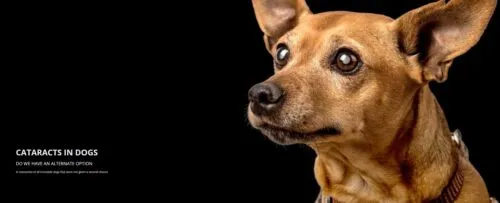
A cataract is an opacity in the lens of a dog’s eye, causing him to have blurry vision. If the cataract is small, it won’t likely disturb the dog’s vision too much, but cataracts must be monitored because the thicker and denser they become, the more likely it is they will lead to blindness.

Cataracts can develop from disease, old age and trauma to the eye, but inherited conditions are the most common cause. Cataracts may be present at birth or develop when a dog is very young-between one and three years of age. A high-incidence of cataracts is also often attributed to diabetes.

There are few types of cataracts in dogs. An immature cataract clouds a greater portion of the lens and can cause some blurred vision. Over time, the entire lens can cloud up and all vision is lost. When this happens, it is known as a mature cataract.

Most cases of cataracts are inherited. For instance, Miniature poodles, American cocker spaniel, miniature schnauzer, golden retrievers, Boston terriers, and Siberian huskies are all predisposed to cataracts.

An untreated cataract may “luxate” or slip from the tissue that holds it in place, freeing it to float around in the eye where it may settle and block natural fluid drainage. This can lead to glaucoma, which can cause permanent blindness. Cataracts may also begin to dissolve after some time, causing deep, painful inflammation in the eye.

REFERENCES

Brian C. Gilger, DVM, MS, Dipl. ACVO. Dipl. ABT
Professor of Ophthalmology, College of Veterinary MedicineNorth Carolina State University, Raleigh, NC

David L. Williams, MA VetMB PhD CertVOphthal MRCVS
St. John’s College, Cambridge. The effect of a topical antioxidant formulation including N-acetyl carnosine on canine cataract: a preliminary study. Veterinary Ophthalmology 2006;9:311-316.

Davidson MG, Nelms SR,
“Diseases of the Canine Lens and Cataract Formation” In: Gelatt KN, Gilger BC,Kern TJ, eds. Veterinary Ophthalmology. 5 ed. Ames: John Wiley & Sons, Inc, 2013;1199-1233.

Kidd PM.
“Bioavailability and activity of phytosome complexes from botanical polyphenols: the silymarin, curcumin, green tea, and grape seed extracts”. Altern Med Rev 2009;14:226-246.

Diagnostic Procedures

The intraocular pressure of the eyes will be assessed to rule out glaucoma. The veterinarian tests the pressure inside the eye using an instrument called a tonometer. Assuming that intraocular pressure is normal, the veterinarian normally will dilate the pupils and use a penlight or other light source to characterize the nature and extent of the cataract and to evaluate for possible concurrent uveitis. Anesthetic drops are normally applied to the eyes before these tests to ensure a painless examination and accurate test results.

Other tests that veterinarians commonly use to diagnose eye conditions include the Schirmer tear test and staining the eye with a fluorescein dye. These two tests are used to check the moisture level of the eye, look for foreign bodies and determine whether damage to the cornea has occurred. Advanced testing may include ocular ultrasound and electroretinography to evaluate the retina and rule out concurrent retinal degeneration. These tests are usually performed if surgery is anticipated. Most of these advanced diagnostic tests typically are performed by a veterinary eye specialist.

dog mature immature cataract eyes

MY DOG HAS CATARACTS. WHAT CAN I DO NOW?

CONTACT YOUR LOCAL VETERINARIAN TODAY

If surgery is recommended by your veterinarian, do not delay. A cataract is a progressive disorder that, if not treated quickly, may lead to blindness in one or both of your dog’s eyes. This is especially the case with diabetes mellitus-related cataracts because they progress very rapidly in dogs. Surgery, however, is often not recommended for dogs with non-hereditary forms of cataract. One modern cataract surgical technique, phacoemulsification, involves the emulsification of the eye’s lens with an ultrasonic handpiece. Once the lens is emulsified and aspirated, aspired fluids are replaced with a balanced salt solution. Also, to prevent extreme farsightedness, an intraocular lens may be implanted during surgery. Phacoemulsification has shown more than a 90 per cent success rate in dogs.

New Hope for Cataracts: NAC (N-Acetylcarnosine) Eye Drops

Carnosine for Vision

Carnosine – a dipeptide consisting of two amino acids (alanine and histidine) connected to each other by a chemical bond called the peptide bond – is one of the most exciting anti-aging nutrients that has recently become widely available. (2) Based on research performed mainly by Russian scientists, it is believed that carnosine is effective both in preventing and treating cataracts. (3-6)

The ability of carnosine to prevent and treat cataracts is believed to be due to its antioxidant properties and its ability to inhibit a chemical process called glycation. Glycation leads to deleterious substances called AGEs (advanced glycation end products). AGEs are chemical complexes that result from common but undesirable reactions between blood sugars, such as glucose, and proteins in many parts of our bodies, including the lenses of our eyes. The sugar-protein complexes become chemically cross-linked and degrade cellular functions. The aptly named AGEs are thought to be an important factor in the aging process.

Carnosine-containing eye drops have demonstrated efficacy in treating a variety of ophthalmic conditions, including corneal diseases, cataracts, glaucoma, and increased intraocular pressure. In 1997, clinical trials with carnosine-containing eye drops were conducted on 109 ophthalmic patients. The results confirmed accelerated healing of corneal erosions, trophic keratitis, post-herpetic epitheliopathy, primary and secondary corneal dystrophy, and bullous keratopathy. (7) Most striking, however, was the ability of carnosine to eliminate existing cataracts. (8)

Carnosine eye drops have been shown to delay vision senescence in humans, being effective in 100 per cent of cases of primary senile cataracts and 80 per cent of cases of mature senile cataracts. Scientists concluded that “carnosine seems to delay the impairment of eyesight with aging, effectively preventing and treating senile cataracts and other age-related diseases.”(9) Carnosine actually restores the proteins in the lens by removing cross-linked carbonyl groups, and is thought to function as a “molecular water pump,” thereby also helping to lower intraocular pressure. (10) In earlier experiments it was demonstrated that applying carnosine to the conjunctiva (the membrane covering the eye) caused a decrease in normal intra-ocular pressure and reduced prostaglandin-induced ocular hypertension (related to glaucoma) in rabbits. (11)

Some scientists believe that carnosine is ineffective if it is metabolized (broken down) by the enzyme, carnosinase. However, studies of corneal transplants in rabbits that were treated with one of the metabolites of carnosine, histidine, indicate that the metabolite itself may be bioactive. Five per cent histidine ointment was applied twice daily to 6 transplants for two months. All six transplants healed and were clear. On the other hand, transplants that were treated with daily applications of one per cent cortisone became opaque, necrotic, and failed to heal. Likewise, transplantation failed completely in six control eyes. (12) This indicates that histidine may be an active portion – if not the active factor – of the carnosine molecule.

Cataract-Dissolving Analog: N-Acetylcarnosine (NAC)

N-acetylcarnitine (NAC), like its parent compound, carnosine, occurs naturally throughout the human body. Both compounds are found primarily in the heart and skeletal muscles (the word carnosine is derived from the Latin word for flesh) and in the brain. Carnosine was discovered in 1900 in Russia, and it is in Russia that most of the recent research on the N-acetyl carnosine derivative has been carried out. (13-15) Research with N-acetylcarnitine, as with carnosine, demonstrates that it is effective not only in preventing cataracts but also in treating them. NAC has been shown to improve vision by partially reversing the development of the cataract, thus increasing the transmissivity of the lens to light.

The structural difference between NAC and carnosine is that one hydrogen atom replaces an acetyl group (CH3CO-), and this substitution occurs at a nitrogen atom. An important chemical difference between carnosine and N-acetylcarnitine is that carnosine is relatively insoluble in lipids (fats and fatty compounds), whereas N-acetylcarnitine is relatively soluble in lipids (as well as in water).

This means that N-acetylcarnitine may pass through the lipid membranes of the corneal and lens cells more easily than carnosine, thereby gaining access more readily to the cells’ interior, which is primarily aqueous. There, the N-acetylcarnitine is gradually broken down to carnosine (and, perhaps, to histidine), which then exerts its beneficial effects.

N-Acetylcarnosine also Reduces Cataracts

In one study, Russian scientists conducted two randomized, double-blind, placebo-controlled trials of 6 months and 24 months duration, with eye drops consisting of a one per cent aqueous solution of NAC administered as two drops twice daily. (16) They treated a total of 49 elderly patients (average age 65) with cataracts ranging in severity from minimal to advanced (but not to the point of requiring surgery); the total number of eyes affected was 76. Using a variety of sophisticated optical techniques, they monitored the condition of cataracts, visual acuity, and glare sensitivity.

The eyes treated with NAC were substantially improved in 6 months: the measured transmissivity of the lenses increased in 42 per cent of the eyes, by 12-50 per cent; in 90 per cent of the eyes, visual acuity improved by 7-100 per cent; and in 89 per cent of the eyes, glare sensitivity improved by 27-100 per cent. These improvements were sustained for the duration of the 24-month trial. In no eyes was any worsening of the condition seen. By contrast, the condition of the untreated eyes in the control group worsened. Visual acuity dropped in 89 per cent of the controls by 17-80 per cent after 24 months.

Another interesting study by the same team also evaluated patients between the ages of 48 and 60, who had various degrees of eyesight impairment, but who did not have the symptoms of cataracts. After a course of treatment ranging from 2 to 6 months, the conclusion was that the eye drops alleviated eye-tiredness and continued to improve eyesight (i.e. there was more clear vision). The subjects reported that the treatment “brightened” and “relaxed” their eyes. This is an important indicator that eye drops have a value both for preventive purposes, as well as medical applications.

Dr. Sara Lam

Share this post to :

Share on facebook
Share on twitter
Share on linkedin
Share on whatsapp