- Bockstahler B, Levine D, Millis D. (2004). Essential Facts of Physiotherapy in Dogs and Cats Rehabilitation and Pain Relief. (BE VetVerlag: Babenhausen, Germany).
- Canapp SO Jr, McLaughlin RM Jr, Hoskinson JJ et al (1999) ‘Scintigraphic evaluation of dogs with acute synovitis after treatment with glucosamine hydrochloride and chondroitin’. Am J Vet Res. Dec;60 (12), 1552 – 1557.
- Carpenter LG, Oulton SA, Piermattei DL. (1996) ‘Femoral head and neck excision in a dog that had previously undergone contralateral hind limb amputation.’ J Am Vet Med Assoc. 208 (5): pp 695 -‐6.
- Davidson JR, Kerwin SC, Millis DL. (2005) ‘Rehabilitation for the orthopedic patient.’ Vet Clin Sm Anim Pract. 35 (6): pp 1357 – 1388.
- DeCamp CE. (1995) ‘Dislocations.’ In Small Animal Orthopedics. Olmstead ML (ed). pp 333 – 359.
- Dickinson R. (2005) ‘A proprioceptive stimulus alters weight bearing in the canine hind limb.’ In Proceedings of the 3rd Annual RVC Veterinary Physiotherapy Conference. (North Mymms, Hatfield, Hertfordshire, UK) pp 26 – 34
- Edge‐Hughes LM. (2002) ‘Therapeutic exercise for the canine patient.’ In Proceeding of the 2nd International Symposium on Rehabilitation and Physical Therapy in Veterinary Medicine. pp 59 -‐ 62. (Knoxville, TN, USA).
- Elliott RP. (1983) The New Dog Steps. (Howell Book House: New York, NY, USA).
- Green, S., Buchbinder, R., Hetrick, S. (2003) Physiotherapy interventions for shoulder pain (Review).
Issue 2. Cochrane Collaboration: The Cochrane Library. - Hamilton S, Millis DL, Taylor RA et al. (2004) ‘Therapeutic exercises.’ In Canine Rehabilitation and Physical Therapy. Millis DL, Levine D & Taylor RA eds. pp 244 – 263. (Saunders: St. Louis, Missouori, USA).
- Hesslink R, Armstrong D, Nagendran MV, et al. (2002) ‘Cetylated fatty acids improve knee function in patients with osteoarthritis.’ J Rheumatol. 29 (8): pp 1708 – 1712.
- Hesslink R, Sprouse S. (2002) ‘The effect of a cetylated fatty acid (CFA) for improving quality of life in canines.’ In Proceedings of the 2nd International Symposium on Rehabilitation and Physical Therapy in Veterinary Medicine. (Knoxville, Tennessee, USA) pp 179.
- Holst S, Lund I, Petersson M et al. (2005) ‘Massage‐like stroking influences plasma levels of gastrointestinal hormones, including insulin, and increases weight gain in male rats.’ Auton Neuro. 120: pp 73 – 79.
- Johnson KA, Hulse DA, Hart RC et al (2001) ‘Effects of an orally administered mixture of chondroitin sulfate, glucosamine hydrochloride and manganese ascorbate on synovial fluid chondroitin sulfate 3B3 and 7D4 epitope in a canine cruciate ligament transaction model of osteoarthritis’. OsteoArthritis and Cartilage. 9, 14 – 21.
- Lephart, S.M., Pincivero D.M., Giraldo J.L. et al. 1997, The role of proprioception in the management and rehabilitation of athletic injuries. Am. J. Sports Med. 25 (1): 130 – 137.
- Levine D. (2006) ‘Physical rehabilitation.’ NAVC Clinicians Brief. 4 (8): pp 51 – 52.
- Michlovitz SL. (1990) ‘Thermal Agents in Rehabilitation .’ (F. A. Davis Company: Philadelphia, PA).
- Miller WH, Scott DW, Wellington JR. (1992) ‘Treatment of dogs with hip arthritis with a fatty acid supplement.’ Canine Pract. 17: pp 6 – 8.
- Moses P. (2006) ‘Module 1. Canine Orthopaedics.’ In Pathological Conditions in Animals II. McGowan, Moses, Malikides (eds). (University of Queensland, Australia).
- Nelson RM, Currier DP. (1987) Clinical Electrotherapy. (Appleton & Lange: Norwalk, CT).
- Ormond AN. (1961) ‘Treatment of hip lameness in the dog by excision of the femoral head.’ Vet Rec. 73: pp 576 – 577.
- Rawson EA, Aronsohn MG, Burk RL. (2005) ‘Simultaneous bilateral femoral head and neck ostectomy for the treatment of canine hip dysplasia.’ J Am Anim Hosp Assoc.41(3): pp 166‐70.
- Saini, N.S., Roy K.S., Bansal P.S. et al. 2002, A preliminary study on the effects of ultrasound therapy on the healing of surgically severed Achilles tendons in five dogs. J. Vet. Med. Assn. 49: 321 – 328.
- Spreull JSA. (1961) ‘Excision arthroplasy as a method of treatment of hip joint diseases in the dog.’ Vet Rec. 73: p 573 – 576.
- Wallace LJ & Olmstead ML. (1995) ‘Disabling conditions of the canine coxofemoral joint.’ In Small Animal Orthopedics. Olmstead ML (ed). pp 361 – 393.
- Witz M, Lepage OM, Lambert C et al. (2001) ‘Brown bear (ursus arctos arctos) femoral head and neck
excision.’ J Zoo Wildlife Med. 32 (4): pp 494 – 499. - Zink, M.C. 1997, Peak Performance ‐ Coaching the Canine Athlete: Canine Sports Productions: Lutherville, MD, USA
PHYSIOTHERAPY | REHABILITATION | HYDROTHERAPY
Relation Of Rehabilitation For Post Surgery
In memories of all immobile dogs that were not given a second chance
What is relation of canine rehabilitation for post surgeries?
Post-operative patients are the most obvious rehabilitation candidates. Just like in people, dogs need more than just strict rest to fully recover from orthopedic procedures. Surgeons have learned that six weeks of strict cage rest after surgery does not result in healthy dogs or happy owners. Rehabilitation helps ease a dog back into normal activities by safely strengthening the muscles around the weakened bones or joints.
Rehabilitation therapy in veterinary medicine is similar to physical therapy in people, but has some significant differences due the variations in the animal’s anatomy and the extreme forces that can be placed on their joints, bones, tendons and muscles.
EFFECTS OF POSTOPERATIVE REHABILITATION
In an independent experiment conducted on 51 client-owned dogs, weighing between 20 and 40 kg (44 to 88 lb) that had RCCL and medial meniscal injury were studied. After removal of the RCCL and complete medial meniscectomy, the stifle joint was stabilized by use of a lateral retinacular stabilization technique.
25 dogs were included in a postoperative rehabilitation group, and 26 dogs were included in an exercise-restricted group. Limb function (peak vertical force [PVF] and vertical impulse [VI]) was determined before surgery and 6 months after surgery, using force platform gait analysis.
Results—Prior to surgery, mean PVF and VI in affected limbs were similar between groups. Six months after surgery, PVF and VI were significantly increased in dogs of both groups. However, PVF and VI in dogs in the rehabilitation group were significantly greater than those of dogs in the exercise-restricted group. At this time, differences in limb function (as measured by PVF and VI) between the repaired and normal limbs were not evident in dogs in the rehabilitation group. Conversely, limb function in the repaired limb of dogs in the exercise-restricted group was still significantly less than that of the normal limb.
References
Darryl L. Millis, MS, DVM, DACVS, DACVSMR, CCRP
Professor of Orthopedic Surgery & Director of Surgical Service
Robin Downing, DVM, MS, DAAPM, DACVSMR, CVPP, CCRP
Diplomate of the American Academy of Pain Management, is a a founder and past-president of the International Veterinary Academy of Pain Management.
Janet B. Van Dyke, DVM
Diplomate American College of Veterinary Sports Medicine and Rehabilitation, CCRT, CEO
Ludovica Dragone, DVM, CCRP
Vice President of VEPRA, Veterinary European of Physical Therapy and Rehabilitation Association.
Andrea L. Henderson, DVM, CCRT, CCRP
Resident, Canine Sports Medicine and Rehabilitation
Steven M.Fox, MS, DVM, MBA, PhD
President Securos. Inc
The Benefits of Post Surgery Rehabilitation and Physiotherapy
After a surgery, rehabilitation therapy can be beneficial for animal companions by
- Exercise intolerance
- Reducing inflammation and pain after surgery
- Promoting early weight bearing
- Strengthening supporting tissues
- Decreasing compensatory muscle, myofascial, and back pain
- Providing mental stimulation for pets who have had restricted activity
- Improving coordination and balanceReversing muscle atrophy
- Promoting weight loss if needed
- Developing structured homework program
- Safely promoting faster return to function
THE ROLE OF REHABILITATION IN POST SURGERY
Physical rehabilitation has become a proven staple in human medicine and doctors now recommend physical rehabilitation following a variety of procedures. These same techniques and modalities are now being applied to veterinary medicine. Canine physical rehabilitation is used to improve the performance and quality of movement for our pets as well as speed healing and provide positive psychological effects.
Following certain surgeries pets may lose up to one third of their muscle mass in a matter of weeks and it may take that same pet more than a year to regain the lost muscle mass. Rehabilitation makes it easier and less stressful for patients to return to functional activities in their day to day life. The prognosis for return to function depends on a great number of variables including whether the pet is a working dog or house pet, as well as the degree of injury and overall body condition.
– Dr. Michael Wolf, DVM, Dr. Med. Vet.Diplomate ACVIM (Neurology), CCRT

UNILATERAL COXOFEMORAL EXCISION ARTHROPASTY
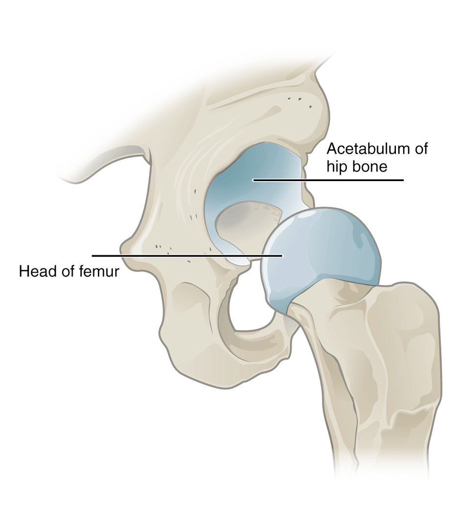
A coxofemoral excision arthroplasty involves the surgical removal of the femoral head and neck. It is a salvage procedure with the goal to eliminate bone to bone contact at the hip and results in the formation of a functional pseudarthrosis (a scar tissue joint).
It was first described as a treatment for dogs with hip dysplasia in 1961. This surgical option is indicated in cases of canine hip dysplasia, Legg-Calve-Perthés disease, non-reparable fractures of the acetabulum or femoral head, osteoarthritis, chronic recurrent hip luxations, osteomyelitisand septic arthritis, failed total hip replacement or villinodular synovitis of the hip joint.
IMPORTANCE OF POST SURGERY PHYSICAL THERAPY
Physical therapy should be started the day after surgery, and incorporate range of motion (ROM) into flexion, extension and abduction. Aggressive analgesic therapy may assist in early ambulation and rapid return to normal function. As well, use of therapeutic modalities can aid in pain relief and soft tissue healing and hence improved functioning.
However, cases that are not referred for physiotherapy immediately post‐op may exhibit severe loss of range of motion and muscle wastage depending on the length of delay before obtaining these services. The goals in treating these cases are to maximize weight bearing, gain ROM or muscle extensibility, strengthen the affected limb, stimulate soft tissue healing, provide pain relief following an aggressive physiotherapy stretching or exercise treatment session, and address proprioception deficits.
Facilitation of Weight bearing
It is imperative that the animal actually use the post‐operative limb, so any safe exercise that encourage limb use can be utilized. Various therapeutic exercise techniques have been described to stimulate weight bearing on any post‐operative limb.
Gain Range of Motion
Regaining hip extension is a one of the most important goals when rehabilitating this type patient. Passive range of motion (PROM) may help to regain extension, however an animal that has been avoiding this movement, the physiotherapist will need to be tricked into extending its hips.
Strengthening
Atrophy of the hind limb muscle mass should be addressed post‐operatively. Various treatment techniques can be used to strengthen the muscles of the hip and thigh. For dogs that are unable to walk, NMES and TENS are often applied.
Pain relief and soft tissue healing
The FHNE surgery may have resulted in adhesions to the distal muscle secondary to tracking of blood from the surgical site and soft tissue damage done at the same time. As well, the techniques listed above may result in mild soft tissue soreness. Therapies that may address this could include modalities and/or massage.
It is well known that healing of soft tissue structures can be accomplished with ultrasound, Low Level Light Therpy (LLLT or also commonly known as Cold Laser), or pulsed electromagnetic field therapy. The tissues surrounding the hip joint as well as the quadriceps and sartorius muscle may benefit from these treatments in regards to the surgically induced trauma. Non‐noxious sensory stimuli reduces blood pressure, changes secretion of corticotropin releasing hormone and increases pain thresholds in rats.
In humans, it produces pain relief and increase the plasma concentration of B‐endorphins. All of these effects could provide comfort to the animal, hence encouraging use and relieving post‐exercise discomfort.
Proprioceptive Retraining
As the limb begins to heal and the dog is utilizing it consistently in gait, the next goal should be to retrain proprioception of the affected leg. Proprioception is the minds awareness of the where the body is in space. Several exercises have been described to address co‐ordination and muscular control of a limb.