
Legg-Calvé-Perthes Disease
This page is for educational purposes only and is not a substitute for veterinary diagnosis or emergency care. Always consult your primary veterinarian or a rehabilitation veterinarian before starting treatment. If your pet cannot walk, has sudden paralysis, severe pain, or breathing difficulty, seek urgent veterinary attention.
What is Legg-Calvé-Perthes Disease?
Also known as: avascular necrosis of the femoral head; aseptic necrosis of the femoral head; Perthes disease; LCPD.
In Legg-Calvé-Perthes disease, blood supply to the developing femoral head is disrupted, leading to bone necrosis, pain, and eventual collapse and remodelling of the head and neck. Toy and small breeds (for example terriers, poodles, miniature breeds) are predisposed; signs usually appear in immature dogs.
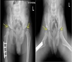
Early radiographs may lag behind clinical pain; serial imaging shows progressive femoral head changes. The condition is distinct from hip dysplasia, though both cause hip pain.
Femoral head and neck ostectomy (FHO) is a common surgical treatment to remove the painful necrotic head; total hip replacement is an option in selected cases. Rehabilitation is critical after FHO to build a functional false joint through early controlled use and progressive strengthening.
Common signs to watch for
Signs vary by severity and by whether your pet is a dog or cat. Owners of dogs often notice:
- Progressive hind-limb lameness in a young small-breed dog
- Pain on hip manipulation; muscle atrophy of the thigh
- Reluctance to jump or play; irritability when the hip is touched
- Shortened stride; weight shifted off the affected limb
- In advanced cases: crepitus and severely reduced hip motion
Causes & contributing factors
- Ischaemia / avascular necrosis of the capital femoral epiphysis
- Genetic predisposition in many small and toy breeds
- Exact vascular trigger often idiopathic
- Not the same disease process as large-breed hip dysplasia
How veterinary rehabilitation helps
After FHO, early rehab encourages protected weight-bearing, range of motion, and rapid rebuilding of gluteal and thigh muscle so the pseudoarthrosis functions well.
Pain-relief modalities, assisted standing, and progressive therapeutic exercise are tailored to the surgical timeline. Hydrotherapy often helps once incisions and soft tissues allow.
Pre-operative rehab may maintain muscle when surgery is planned. Medical management alone is less common once collapse is painful — your vet decides.
Rehabilitation plans at RehabVet are individualised after a veterinary assessment. We coordinate with your primary vet when imaging, medication, or surgery is part of the overall plan.
Modalities & services commonly used at RehabVet
Depending on your pet’s examination findings, comfort, and goals, a plan may include one or more of the following:
Expected rehabilitation goals
Goals are set for the individual patient. Typical aims may include (not guarantees — outcomes vary):
- Relieve hip pain from necrotic femoral head disease
- After FHO: establish comfortable, strong false-joint function
- Restore weight-bearing and hind-limb muscle mass
- Improve jumping, stairs, and play confidence appropriate to the dog
- Guide owners through home exercises and activity progression
We do not publish invented success percentages. Progress is tracked clinically (gait, strength, range of motion, pain behaviours, and home function) and plans are adjusted over time.
When to seek veterinary care
- Persistent hind-limb limp in a young small-breed dog
- Pain when the hip is handled; rapid muscle loss
- Sudden refusal to use a hind limb
- Post-FHO concerns: incision issues or reluctance to begin using the leg when advised
No. Perthes disease is avascular necrosis of the femoral head in young small dogs. Hip dysplasia is a developmental incongruity/laxity problem more often discussed in larger breeds — though both can end in OA and may be treated with FHO in some cases.
FHO relies on soft tissues and muscle to create a comfortable false joint. Early, guided use and strengthening strongly influence functional results.
Bilateral disease can occur. Your veterinarian will assess both hips even if only one limb appears lame.
Related reading & patient stories
Book a rehabilitation assessment
If your pet has been diagnosed with Legg-Calvé-Perthes, or you are noticing mobility changes, our team can assess and design a multimodal rehab plan.
Educational content only — not a diagnosis. For emergencies, contact your nearest veterinary hospital.
Related Conditions
- ConditionAngular Limb DeformityAngular limb deformity describes abnormal angulation or rotation of a limb from asymmetric growth-plate function or trauma, altering joint alignment and gait in dogs.Learn more
- ConditionElbow DysplasiaElbow dysplasia is a group of developmental elbow disorders — including FMCP, UAP, OCD, and incongruity — that cause forelimb pain and progressive osteoarthritis, mainly in dogs.Learn more
- ConditionHip DysplasiaBunny-hopping, struggling to stand, sore after exercise? Many dysplastic dogs are managed well without surgery. RehabVet builds the muscle that supports the joint and keeps your dog moving comfortably.Learn more