
Lumbosacral Stenosis
This page is for educational purposes only and is not a substitute for veterinary diagnosis or emergency care. Always consult your primary veterinarian or a rehabilitation veterinarian before starting treatment. If your pet cannot walk, has sudden paralysis, severe pain, or breathing difficulty, seek urgent veterinary attention.
What is Lumbosacral Stenosis?
Also known as: cauda equina syndrome; degenerative lumbosacral stenosis; LS disease; lumbosacral disease.
The lumbosacral junction (typically L7–S1 in dogs) is a high-motion, high-load segment. Degenerative changes — disc protrusion, ligament hypertrophy, bone remodelling, or instability — can narrow the canal or foramina and irritate the cauda equina nerve roots. Working and large-breed dogs are frequently discussed, but any dog can be affected.
Owners often report difficulty rising, pain when the tail is lifted, reluctance to jump into the car, pelvic-limb lameness that may shift sides, or urinary/faecal changes in more advanced cases. Orthopaedic hip disease can coexist and confuse the picture; careful veterinary examination and imaging guide diagnosis.
Rehabilitation complements medical management and, when surgery is performed, post-operative recovery. Goals include pain reduction, core and pelvic stabilisation, nerve-glide friendly movement, and graded return to activity without provoking flare-ups.
What are the signs of Lumbosacral Stenosis?
Signs vary by severity and by whether your pet is a dog or cat. Owners of dogs often notice:
- Lower-back pain; sensitivity over the lumbosacral junction
- Reluctance to jump, climb, or extend the pelvic limbs
- Pelvic-limb weakness, scuffing, or intermittent lameness
- Chewing at the flank, hips, or tail base; restlessness when lying down
- Tail carriage changes or pain on tail manipulation
- In advanced cases: urinary or faecal incontinence, or proprioceptive deficits
What causes Lumbosacral Stenosis?
- Degenerative disc disease and soft-tissue hypertrophy at L7–S1
- Instability or malalignment of the lumbosacral junction
- Congenital stenosis in some individuals
- Repetitive loading in working, sporting, or overweight dogs
- Concurrent orthopaedic disease increasing compensatory spinal stress
How can veterinary rehabilitation help with Lumbosacral Stenosis?
Rehab programmes emphasise pain control strategies, gentle mobilisation within comfort, and progressive core and hip strengthening to improve dynamic stability around the junction.
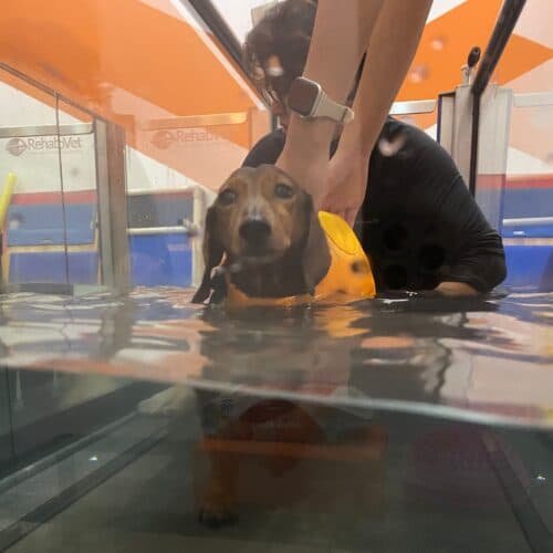
Therapists teach activity modification — avoiding ballistic jumping while rebuilding controlled strength. Modalities and manual therapy may reduce protective muscle spasm. Hydrotherapy can provide low-impact conditioning when pain allows.
Post-surgical patients follow surgeon-specific protocols with staged loading and scar/soft-tissue care.
Rehabilitation plans at RehabVet are individualised after a veterinary assessment. We coordinate with your primary vet when imaging, medication, or surgery is part of the overall plan.
Which therapies may be used for Lumbosacral Stenosis?
Depending on your pet’s examination findings, comfort, and goals, a plan may include one or more of the following:
What are the rehabilitation goals for Lumbosacral Stenosis?
Goals are set for the individual patient. Typical aims may include (not guarantees — outcomes vary):
- Reduce lumbosacral pain and nerve-root irritability
- Improve pelvic-limb strength and proprioception
- Restore safer jumping and rising mechanics where appropriate
- Address compensatory patterns in hips, stifles, and thoracic spine
- Support durable return to daily or working tasks under guidance
We do not publish invented success percentages. Progress is tracked clinically (gait, strength, range of motion, pain behaviours, and home function) and plans are adjusted over time.
When should you seek veterinary care for Lumbosacral Stenosis?
- Persistent lower-back pain, yelping when rising, or refusal to jump
- Progressive pelvic-limb weakness or knuckling
- Changes in urination or defecation
- Acute severe pain or inability to walk — urgent veterinary care
No, but they often coexist. Hip dysplasia or osteoarthritis can mimic or compound LS signs. A veterinary exam (sometimes with imaging of both regions) is needed to prioritise treatment.
Some dogs are managed successfully with multimodal medical care and rehab; others need surgical decompression or stabilisation. Decision-making rests with your veterinarian or surgeon based on neurological deficits, pain, and imaging — not on marketing claims.
During flare-ups, high-impact jumping is usually restricted. Ramps, controlled strength work, and gradual return are safer. Your rehab team will tailor restrictions to your dog’s stage of recovery.
Related reading & patient stories
Book a rehabilitation assessment
If your pet has been diagnosed with LS disease, or you are noticing mobility changes, our team can assess and design a multimodal rehab plan.
Educational content only — not a diagnosis. For emergencies, contact your nearest veterinary hospital.
Related Conditions
- ConditionDiscospondylitisDiscospondylitis is infection of an intervertebral disc and neighbouring vertebral endplates, causing spinal pain, stiffness, fever, and sometimes neurological deficits in dogs (rarely cats).Learn more
- ConditionIntervertebral Disc Disease (IVDD)Veterinary rehabilitation for dogs with IVDD in Singapore — pain relief, neuro-recovery and safe mobility work after disc disease, coordinated with your vet.Learn more
- ConditionWobbler SyndromeWobbler syndrome (cervical spondylomyelopathy) refers to cervical spinal cord compression in dogs, producing a characteristic wobbly gait, neck pain, and progressive neurological deficits.Learn more